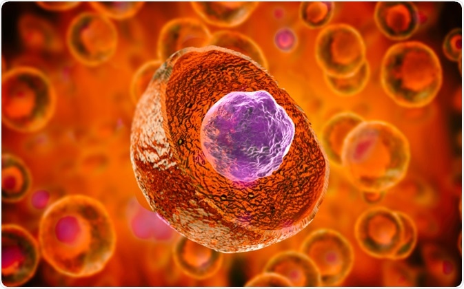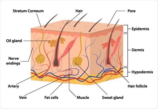
The skin is the largest organ in the body and contains several different types of stem cells. These cells maintain the balance between the production and turnover of the layers of the skin.

Giovanni Cancemi | Shutterstock
Stem cell niches in the skin
The epidermis (outer layer of the skin) has three stem cell niches. The first two are the bulge region of the hair follicle and the basal layer of the interfollicular epidermis (IFE). The final stem cell niche is unknown, and belongs to the melanocyte stem cells.
Stem cells in the hair follicle
In neonatal mice, the bulge SCs repopulate the epidermis and provide a flow of proliferating keratinocytes to the epidermis in both neonatal and adult mice.
In adult mice, the bulge SC only repopulate the epithelium in response to wounding and not homeostasis. However, studies show that stem cells derived from the interfollicular epithelium, rather than the bulge stem cells provided the epithelial cells necessary for wound healing.
Activating bulge stem cells
Signaling molecules, such as Wnt, Notch, and BMP, from the bulge niche normally keep hair follicle stem cells quiescent. These are released by nearby epithelial and mesenchymal cells, which control hair follicle morphogenesis and the formation of the dermal papilla
The BMP signaling pathway controls the proliferation and differentiation of dermal papilla cells at the base of the hair follicle to induce hair follicle growth. The BMP pathway is therefore responsible for stem cell activation in the hair follicle niche. This signaling pathway initiates a cascade of epithelial-mesenchymal interactions which play a key role in the hair cycle, and occur not only during development but throughout a human's lifetime.

NeutronStar8 | Shutterstock
Stem cells of the interfollicular epidermis (IFE)
IFE SCs usually only contribute to the IFE and not hair follicles. However, in cases of severe injury, they can participate in de novo hair follicle regeneration involving the Wnt signaling pathway, as seen in embryonic development. The discovery of Wnt signaling here further demonstrated the role of mesenchymal-epithelial interactions in the formation of the hair follicles.
Melanocyte stem cells
Melanocyte stem cells maintain the population of melanocytes in the skin. These cells produce melanin in response to UV exposure, which protects the DNA of keratinocytes in the outer most layer of the skin from damage. The location of these stem cells is currently unknown.
Functions of stem cells in the skin
The epidermis of the skin and the hair follicles provide a protective barrier against microbes, injury, and desiccation of underlying cells. Skin cells are continually sloughed, so the integrity of this barrier is entirely dependent on regular cell renewal. This homeostatic function is carried out by skin stem cells that reside in the epidermis, the hair follicle, and sebaceous glands, which proliferate in response to external signals.
Sources
- https://www.ncbi.nlm.nih.gov/pmc/articles/PMC2213592/
- https://www.ncbi.nlm.nih.gov/pmc/articles/PMC3842871/
- https://www.sciencedirect.com/science/article/pii/S1021949817301874
Last Updated: Dec 20, 2018

Written by
Hidaya Aliouche
Hidaya is a science communications enthusiast who has recently graduated and is embarking on a career in the science and medical copywriting. She has a B.Sc. in Biochemistry from The University of Manchester. She is passionate about writing and is particularly interested in microbiology, immunology, and biochemistry.
Source: Read Full Article